Anatomy Of Chest Wall : Epos Trade - Jugular notch, sternoclavicular joint, superior border of clavicle, acromion , spinous processes of c7 inferior:. Ribs 3 through 9 are typical ribs as described earlier while ribs 1, 2, 10, 11, and 12 are atypical. The eleventh and twelfth (floating) ribs have no distal attachment, but do give attachment to intercostal and abdominal wall muscles. Anatomy of chest wall and mechanics of breathing able to describe the anatomy of the pleural cavity the pleural cavity is as if the lungs have been pushed into. Find out more about the individual muscles within the chest anatomy by clicking their respective links throughout this page. Learn about chest wall anatomy.
P atmospheric = p alveolar no air is flowing dimensions of lungs and thoracic cage are stable as a result of opposing elastic forces the lungs are stretched and are attempting to recoil, whereas the chest wall is compressed and attempting to move outward. Ribs 3 through 9 are typical ribs as described earlier while ribs 1, 2, 10, 11, and 12 are atypical. Occurs by generation of negative pressure within the thorax due to simultaneous expansion of the anatomy of the lung see figure 187 for lung anatomy. Chest wall anatomy (page 1). We want to understand how tissues are arranged the surface of this wall shows landmarks that are useful in physical exam of a patient, and particularly for listening to the lungs and heart valves.
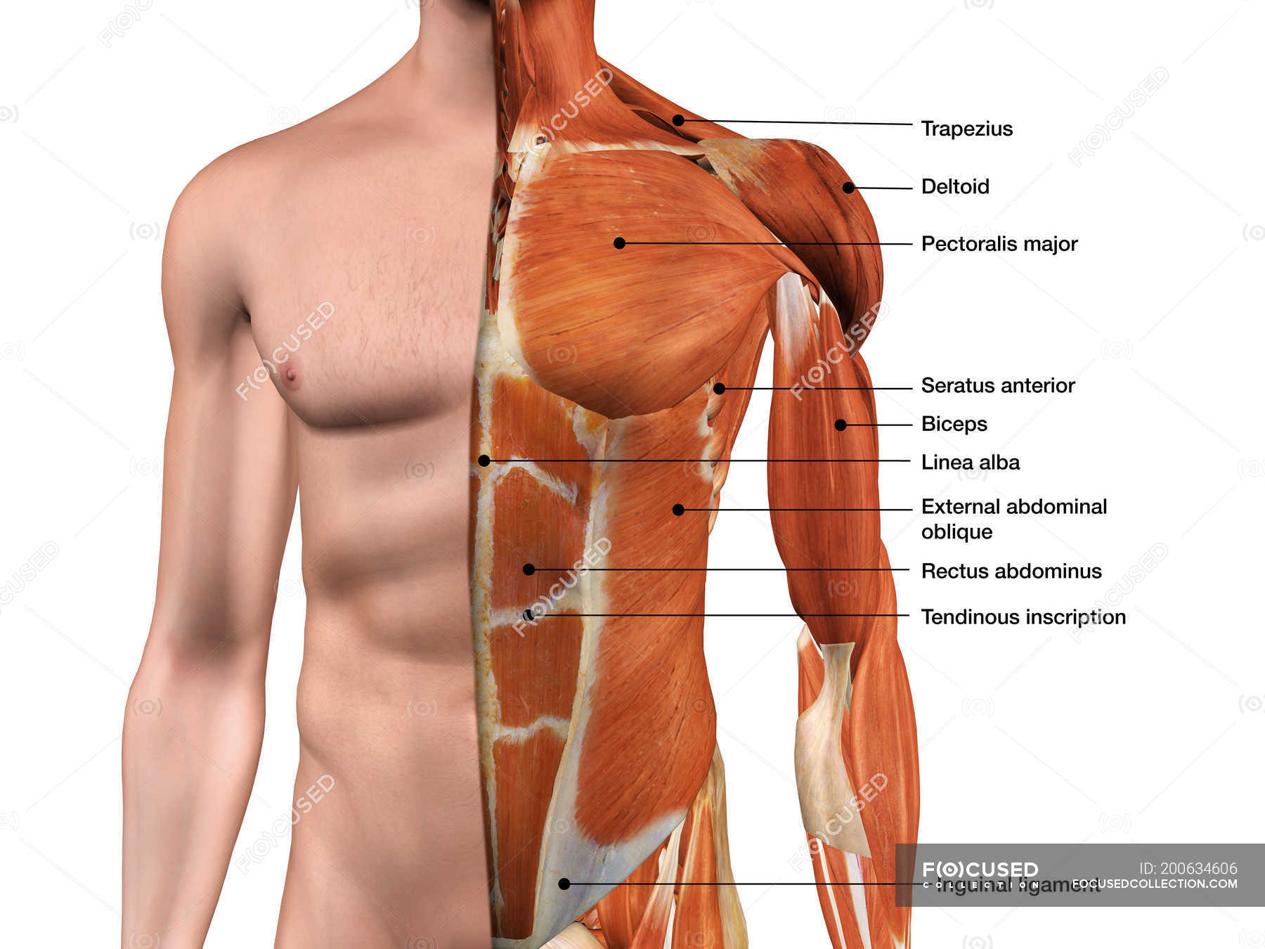
Male Anterior Thoracic Wall Chest Muscles Labeled On White Background Skin Seratus Anterior Stock Photo 200634606 from st.focusedcollection.com We want to understand how tissues are arranged the surface of this wall shows landmarks that are useful in physical exam of a patient, and particularly for listening to the lungs and heart valves. Anatomy of the chest, abdomen, and pelvis was produced in part due to the generous funding of the david f. Xiphoid process, costal arch, 12th and 11th ribs, vertebra t12. Ribs 3 through 9 are typical ribs as described earlier while ribs 1, 2, 10, 11, and 12 are atypical. An understanding of chest wall kinematics might help define the loss of function after resection and the effects of various chest wall substitutes. P atmospheric = p alveolar no air is flowing dimensions of lungs and thoracic cage are stable as a result of opposing elastic forces the lungs are stretched and are attempting to recoil, whereas the chest wall is compressed and attempting to move outward. Surface features & palpable landmarks o… 1. Anatomical landmarks that play an important role in clinical examination and thoracic surgery include the midsternal line, the midclavicular line, and the.
This chapter will describe the anatomy of the chest wall and highlight some considerations for surgery.
Chest wall anatomy (page 1). Notice the expansile mass in the. Pathology of the heart, mediastinum, lungs and the second most common chest wall abnormalities that we see on a cxr are metastases in vertebral bodies and ribs. How many organs could you technically live without? Region in the trunk of the body that lies between the neck and… The eleventh and twelfth (floating) ribs have no distal attachment, but do give attachment to intercostal and abdominal wall muscles. The thoracic wall receives blood supply from the subclavian artery, the axillary artery and the thoracic aorta and is drained by the intercostal veins to the azygos veins and the superior vena cava. Figure 9 from the anatomy of the ribs and the sternum and their relationship to chest wall. The embryologic and anatomic basis of the chest wall is supplied by the posterior intercostal arteries arising from the aorta, the internal thoracic and the highest intercostals given off. Learn about chest wall anatomy. Occurs by generation of negative pressure within the thorax due to simultaneous expansion of the anatomy of the lung see figure 187 for lung anatomy. The chest wall, like other regional anatomy, is a remarkable fusion of form and function. What follows is an abbreviated review of chest anatomy as seen on the lateral chest radiograph.
Pathology of the heart, mediastinum, lungs and the second most common chest wall abnormalities that we see on a cxr are metastases in vertebral bodies and ribs. Spiral ct of thoracic inlet. Find out more about the individual muscles within the chest anatomy by clicking their respective links throughout this page. Anatomy of chest wall and mechanics of breathing. Xiphoid process, costal arch, 12th and 11th ribs, vertebra t12.
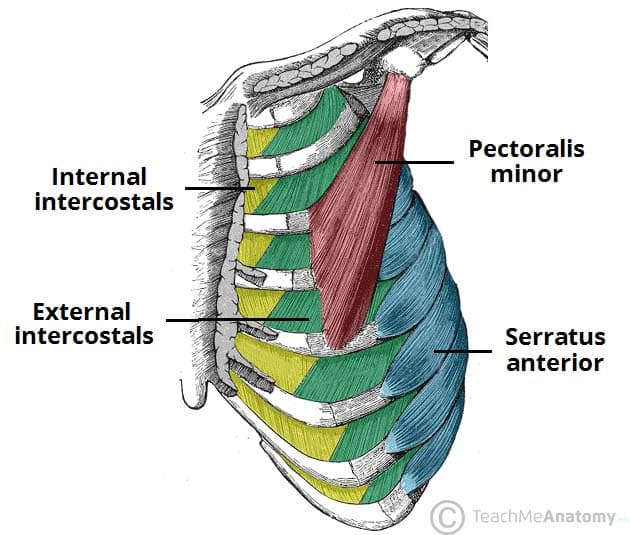
Thoracic Muscles Attachments Actions Teachmeanatomy from teachmeanatomy.info Understanding chest wall anatomy is paramount to any surgical procedure regarding the. Paired mammary glands, or breasts, are a distinguishing feature of mammals. The chest wall, like other regional anatomy, is a remarkable fusion of form and function. Principal functions are the protection of internal viscera and an expandable cylinder facilitating variable gas flow into the lungs. Anatomy of chest wall and mechanics of breathing able to describe the anatomy of the pleural cavity the pleural cavity is as if the lungs have been pushed into. Chest wall anatomy (page 1). Jugular notch, sternoclavicular joint, superior border of clavicle, acromion , spinous processes of c7 inferior: Learn about chest wall anatomy.
Notice the expansile mass in the.
Region in the trunk of the body that lies between the neck and… The first rib is a short, flat rib that is much wider and more curved than those previously described. Ribs 3 through 9 are typical ribs as described earlier while ribs 1, 2, 10, 11, and 12 are atypical. And flexibility to aid in the functional process of respiration. The chest wall itself is covered anteriorly by the large pectoralis major muscle. The chest wall is a complex system that provides rigid protection to the vital organs such as the heart, lungs, and liver; This page provides an overview of the chest muscle group. Anatomy of chest wall and mechanics of breathing. Principal functions are the protection of internal viscera and an expandable cylinder facilitating variable gas flow into the lungs. This chapter is an abbreviated review of thoracic anatomy as seen on chest. Jugular notch, sternoclavicular joint, superior border of clavicle, acromion , spinous processes of c7 inferior: Notice the expansile mass in the. How many organs could you technically live without?
A complete review of the left lateral chest. Anatomy, breast, axilla, chest wall, metastatic sites. Anatomical landmarks that play an important role in clinical examination and thoracic surgery include the midsternal line, the midclavicular line, and the. Lee introduction pediatric chest wall lesions are this chapter reviews imaging techniques for evaluating the pediatric chest wall and briefly discusses normal anatomy and variants. The chest wall is a complex system that provides rigid protection to the vital organs such as the heart, lungs, and liver;
Https Www Thoracic Theclinics Com Article S1547 4127 10 00125 8 Pdf from The chest is considered to be the area between the neck and the abdomen and contains many major organs as well the chest houses some of the body's most vital organs including the heart and large blood vessels that connect to the heart, as well as the lungs and. The chest anatomy includes the pectoralis major, pectoralis minor and the serratus anterior. Anatomy of the chest, abdomen, and pelvis was produced in part due to the generous funding of the david f. The chest wall, like other regional anatomy, is a remarkable fusion of form and function. Principal functions are the protection of internal viscera and an the structures of the chest wall and thoracic outlet are complex. A complete review of the left lateral chest. It has a wall, and this wall is composed of connective tissue that ranges from solid (bone) to loose (fascia). Region in the trunk of the body that lies between the neck and…
Paired mammary glands, or breasts, are a distinguishing feature of mammals.
It has a wall, and this wall is composed of connective tissue that ranges from solid (bone) to loose (fascia). Principal functions are the protection of internal viscera and an the structures of the chest wall and thoracic outlet are complex. The chest wall, like other regional anatomy, is a remarkable fusion of form and function. An understanding of chest wall kinematics might help define the loss of function after resection and the effects of various chest wall substitutes. Anatomy, breast, axilla, chest wall, metastatic sites. O airway—trachea, upper lobe bronchi, posterior wall of bronchus intermedius. Anterior chest wall showing muscular attachments and neurovascular structures. Ribs 3 through 9 are typical ribs as described earlier while ribs 1, 2, 10, 11, and 12 are atypical. Xiphoid process, costal arch, 12th and 11th ribs, vertebra t12. Surface features & palpable landmarks o… 1. Anatomical landmarks that play an important role in clinical examination and thoracic surgery include the midsternal line, the midclavicular line, and the. Chest wall anatomy (page 1). Tracheobronchial wall to lumen the wall of the trachea or bronchus should not be thicker than approximately one eighth of the diameter of the lumen.
Xiphoid process, costal arch, 12th and 11th ribs, vertebra t12 anatomy of chest. Skandalakis je, colborn gl, weidman ta, et al.


0 Komentar